


Dysplastic degenerative disease that causes destruction of the hip and has the chronic character of the current. It is more common in the older age groups. Often sick women than men.
The beginning of the disease progressive, develops slowly. It may affect one joint or both. It is the most common type of osteoarthritis.
Why the disease develops?
Dysplastic in some patients is accompanied by the natural process of aging of the organism and is the dystrophy of the tissues of the hip. In its appearance influencing factors:
- reduction of the nutrition of the tissues;
- a congenital defect of the hip, in particular, dysplasia;
- a pelvic trauma;
- post-infectious dysplastic;
- avascular necrosis of the head of the hip;
- perthes disease (osteochondropathy).
Unfortunately, determine the cause of the disease is not always can and pathology of the hip is called idiopathic coxarthrosis - that is to say, the causes of which are not installed. This is a permanent incentive to the research problem. Work is being carried out scientists in this field and the doctors came to the conclusion that a higher risk of coxarthrosis is observed in the following patients of patients:
- The propensity of hereditary pathology. Patients whose parents suffered from diseases of cartilage and bone, in most cases, are also going to have similar problems;
- The excess of weight. A large mass of a body is the load on the joints, and you regularly work to machine the work;
- The types of infraction, diabetes mellitus. This leads to poor admission of oxygen and nutrients in the tissue of the joint, so that they are losing their properties.
Knowing the main risk factors of the disease, it is possible to plan the preventive measures for its prevention.
How to recognize the pathology of the hip?
The symptoms coxarthrosis depends on the anatomical features of the locomotor system, the causes of the pathology and the stage of the process. Let's look at the main clinical manifestations:
- the pain of the joint;
- the irradiacin of pain in the knee, the hip, the adductors;
- the stiffness of movement;
- the limited mobility;
- the rape of a foot, lameness;
- the decrease of the mass of the muscles of the hip;
- the shortening of the injured limb.
The clinical picture corresponds to internal changes in the tissues of the joint. The symptoms increase gradually and in the early stages of the patient is not given the due attention. This is dangerous, because it is in the beginning of the process of treatment of the cause more effect.
Clinical and radiological measurement coxarthrosis
Below is a list of the symptoms of the disease, specific to each grade level.
- 1 degree. The patient feels regular pain and discomfort. The unpleasant sensations bother you after you do exercise, a lot of the situation of a static posture. The pain is located in the area of the joint and passes it after the holidays. At this stage of the process does not break the gear and no shortening of the leg. Changes visible on x - ray- narrowed joint cleft, in which there are osteophytes (bone expansion).
- 2 degree. Increases the intensity of the pain, which can appear during the resting and radiating in adjacent areas of the body. It appears the limp after that the man was going for a long time or surge. Is limited to the amount of movement in the joint. In parallel develop the changes x-ray: moves the head of the femur, osteophytes grow on the inside and the outside of the edges of the acetabulum.
- 3 phase. The pain acquires a permanent character, that appears in the day and the night. It is much more difficult to walk, appears the constant lameness. It drastically reduces the propulsion of the function, atrophy of the muscles of the legs. change in the muscle tissue makes the walk a little "pulled", and is made shorter. This leads to the deformation of the posture and the curvature of the body. The x-ray at this stage of the process: the total narrowing of the slit between the surfaces of the joint, the deformation of the femoral head, the significant increase in osteophytes.
Program diagnostic of the disease
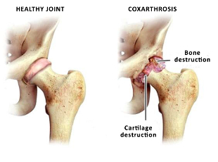
The main method of diagnosis - x-ray. With your help we can determine the presence of the disease and its state. In the x-ray analysis of the structure of the joint in the subject of narrowing of the joint cleft, the osteophytes, the destruction of the head of the hip bone.
If there is a need in the study of the state of the soft tissues, carries out magnetic resonance tomography. Allows you to query the state of the cartilage of the plots of the joint as well as muscles of the hip area.
The modern methods and the direction of treatment of coxarthrosis of the hip
The treatment of coxarthrosis can be conservative and surgical. The treatment of coxarthrosis directed to the achievement of the following objectives:
- the reduction of pain manifestations;
- the recovery of motor activity;
- the rehabilitation and recovery of work capacity;
- prevention of complications;
- the improvement of the quality of life of the patient.
The start of the treatment consists in the modification of risk factors. To do this, the doctor recommends the following activities:
- the normalization of the mass of the body;
- the rejection of bad habits;
- nutrition;
- the standardization of the physical activity;
- a balanced regime of drinking;
- the healthy sleep.
The conservative treatment are distinguished: - drug and non-drug. The pharmacological treatment includes nonsteroidal anti-inflammatory drugs, analgesics, chondroprotectors. Reduces the inflammatory process in the tissues of the joint, remove the swelling and pain, restore range of motion and improve the conditions of the cartilage tissue.
Non-drug treatment includes, among other things, the massage of the affected area. This stimulates the functioning of the muscles, to resist their dystrophy and the prevention of the shortening of the limb. A complete and professional massage stimulates the blood flow in the area of the joint, and this, in turn, leads to the normalization of metabolism in the tissues. Please note that massage is not always helpful when coxarthrosis - is performed only between the acute episodes and in some stages of the process. Assigned to your health care provider may be the doctor can recommend the massage techniques, the multiplicity of procedures and the duration of the course.
Requirement for the treatment, therapeutic exercise. Is the prevention of contractures and progression of the disease. The exercise that is done every day, only have effect. Gymnastics is selected individually and is assigned a doctor. The exercises improve the general well-being, reduce the risk of emotional disorders, strengthen the body's strength.
Physical therapy is another method that is applied in coxarthrosis. This may be of mud, therapeutic baths and shower, magnet. Applies to electricity, and phonophoresis with medicinal substances.
If these treatments have not produced the effect or used outside of the time - it is necessary to surgical treatment.
Early intervention in coxarthrosis
The surgical treatment is applied to the ineffectiveness of conservative methods. This is especially true with the subsequent diagnosis. The modern operational methodologies and quality of equipment operating to restore the structure and function of the joint, return to the person the amount of normal movement and quality of life. The most effective method for the surgical treatment is the replacement of the joint.
The indications to early intervention are:
- coxarthrosis 2-3 degrees;
- the lack of effect of the therapy;
- total restriction of movements to walk.
Contraindications, which do not allow to perform the operation:
- decompensated the state of the kidneys, heart, liver;
- mental illness;
- the acute phase of the inflammatory process in the body.
Precisely this is the preoperative diagnosis. However, if there is the possibility of adjusting the state of the patient is prepared for the operation and then carried out the intervention.
The operation consists in the removal of tissues and the installation of the prosthesis. There are different models of these implants. Vary the methods of fixation in the bone – cement and non-cemented, the material of the implant. About all the features of a stent and the complexities of the intervention, you can obtain information on the consultation of your doctor.
The recovery period after the surgery
From the first day after the surgery, is performed the rehabilitation under the supervision of a doctor. First, is the execution of passive movements, and then load is gradually increasing. Walking in the first time is allowed only with crutches, it allows the seat and bend over.
Naturally, in the first time after the surgery, there are limitations on the loads. Do not be afraid - because without the operation of these restrictions have been retained until the end of life. The decrease of physical activity after the surgery, the treatment is necessary to improve the position of a stent-graft, the restoration of the integrity of the bones, the healing of wounds. In the course of 2 months should be excluded from sports, physical activities in the joint, the long hike and some types of exercises. After the recovery is complete the person returns to full life, you can practice sports and active leisure activities.
The terms of service of a stent: the majority of the companies indicates the survival rate of about 90% for the observation time of up to 15 years.